

Where In The Body Would You Be Most Likely To Find Secretory Cells Shaped Like Dice?
The Tissue Level of Organization
Epithelial Tissue
Learning Objectives
By the end of this section, you will be able to:
- Explain the structure and role of epithelial tissue
- Distinguish between tight junctions, anchoring junctions, and gap junctions
- Distinguish between uncomplicated epithelia and stratified epithelia, besides every bit between squamous, cuboidal, and columnar epithelia
- Describe the structure and part of endocrine and exocrine glands and their corresponding secretions
Most epithelial tissues are essentially big sheets of cells covering all the surfaces of the body exposed to the outside earth and lining the exterior of organs. Epithelium also forms much of the glandular tissue of the torso. Skin is not the only expanse of the torso exposed to the exterior. Other areas include the airways, the digestive tract, besides as the urinary and reproductive systems, all of which are lined by an epithelium. Hollow organs and body cavities that do not connect to the exterior of the torso, which includes, blood vessels and serous membranes, are lined past endothelium (plural = endothelia), which is a type of epithelium.
Epithelial cells derive from all three major embryonic layers. The epithelia lining the skin, parts of the mouth and nose, and the anus develop from the ectoderm. Cells lining the airways and most of the digestive system originate in the endoderm. The epithelium that lines vessels in the lymphatic and cardiovascular arrangement derives from the mesoderm and is called an endothelium.
All epithelia share some important structural and functional features. This tissue is highly cellular, with piddling or no extracellular material present between cells. Adjoining cells form a specialized intercellular connection between their jail cell membranes called a cell junction. The epithelial cells exhibit polarity with differences in structure and function between the exposed or upmost facing surface of the cell and the basal surface shut to the underlying body structures. The basal lamina, a mixture of glycoproteins and collagen, provides an zipper site for the epithelium, separating it from underlying connective tissue. The basal lamina attaches to a reticular lamina, which is secreted past the underlying connective tissue, forming a basement membrane that helps concur it all together.
Epithelial tissues are nearly completely avascular. For instance, no blood vessels cantankerous the basement membrane to enter the tissue, and nutrients must come up by diffusion or assimilation from underlying tissues or the surface. Many epithelial tissues are capable of speedily replacing damaged and dead cells. Sloughing off of damaged or expressionless cells is a characteristic of surface epithelium and allows our airways and digestive tracts to rapidly supersede damaged cells with new cells.
Generalized Functions of Epithelial Tissue
Epithelial tissues provide the trunk'southward showtime line of protection from physical, chemical, and biological wearable and tear. The cells of an epithelium act as gatekeepers of the body controlling permeability and allowing selective transfer of materials across a concrete barrier. All substances that enter the body must cross an epithelium. Some epithelia often include structural features that permit the selective ship of molecules and ions across their prison cell membranes.
Many epithelial cells are capable of secretion and release mucous and specific chemic compounds onto their apical surfaces. The epithelium of the pocket-sized intestine releases digestive enzymes, for instance. Cells lining the respiratory tract secrete mucous that traps incoming microorganisms and particles. A glandular epithelium contains many secretory cells.
The Epithelial Prison cell
Epithelial cells are typically characterized by the polarized distribution of organelles and membrane-bound proteins between their basal and apical surfaces. Particular structures institute in some epithelial cells are an adaptation to specific functions. Certain organelles are segregated to the basal sides, whereas other organelles and extensions, such every bit cilia, when present, are on the apical surface.
Cilia are microscopic extensions of the apical jail cell membrane that are supported by microtubules. They beat in unison and move fluids as well as trapped particles. Ciliated epithelium lines the ventricles of the brain where information technology helps circulate the cerebrospinal fluid. The ciliated epithelium of your airway forms a mucociliary escalator that sweeps particles of dust and pathogens trapped in the secreted mucous toward the throat. It is chosen an escalator because it continuously pushes mucous with trapped particles upwardly. In contrast, nasal cilia sweep the mucous blanket downwardly towards your pharynx. In both cases, the transported materials are usually swallowed, and end up in the acidic surround of your stomach.
Cell to Prison cell Junctions
Cells of epithelia are closely connected and are not separated by intracellular material. Three bones types of connections allow varying degrees of interaction between the cells: tight junctions, anchoring junctions, and gap junctions ((Effigy)).
Types of Cell Junctions
The three basic types of prison cell-to-cell junctions are tight junctions, gap junctions, and anchoring junctions.
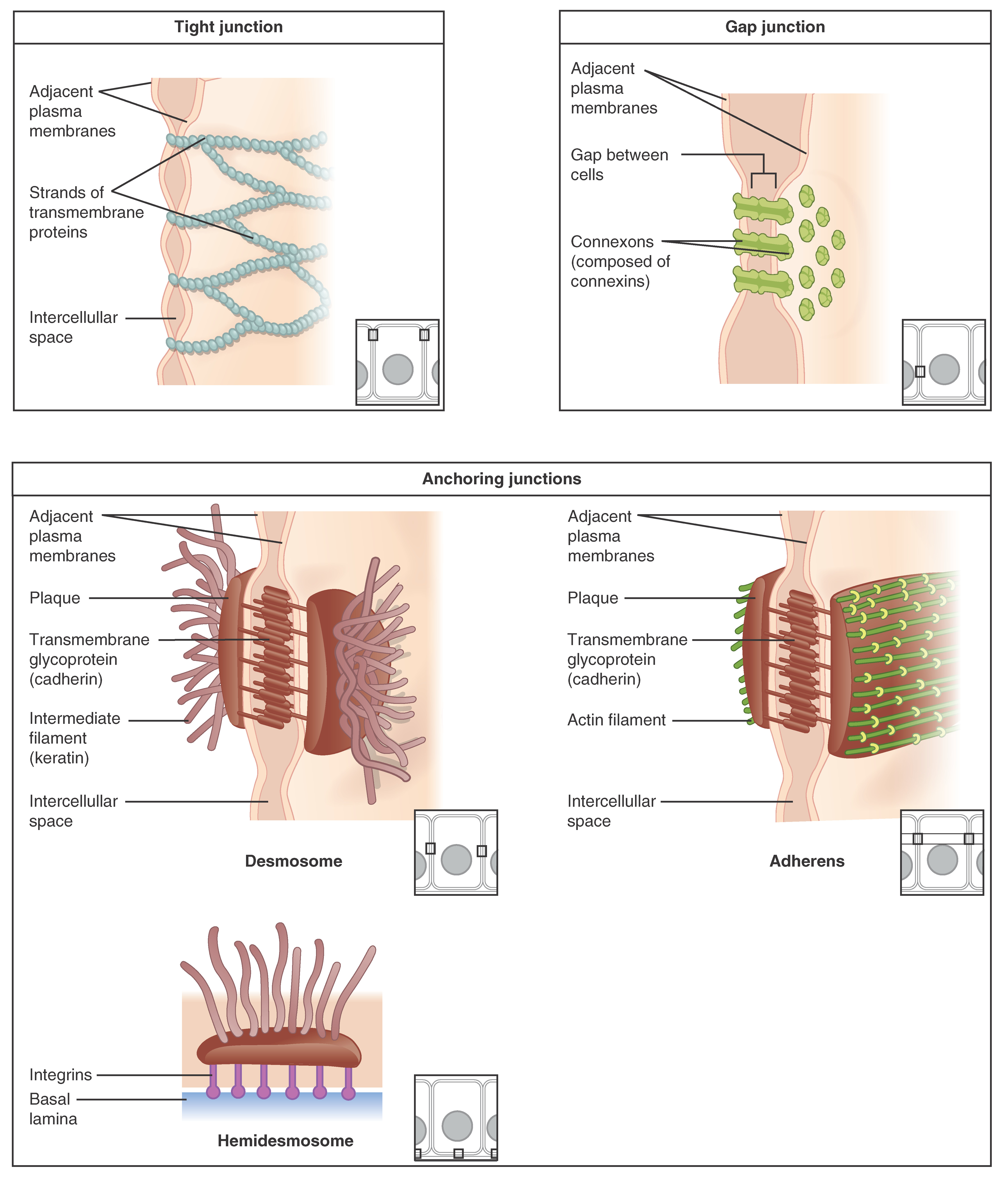
At one end of the spectrum is the tight junction, which separates the cells into apical and basal compartments. When two adjacent epithelial cells form a tight junction, there is no extracellular infinite betwixt them and the motion of substances through the extracellular space between the cells is blocked. This enables the epithelia to act equally selective barriers. An anchoring junction includes several types of prison cell junctions that help stabilize epithelial tissues. Anchoring junctions are mutual on the lateral and basal surfaces of cells where they provide strong and flexible connections. In that location are three types of anchoring junctions: desmosomes, hemidesmosomes, and adherens. Desmosomes occur in patches on the membranes of cells. The patches are structural proteins on the inner surface of the cell'southward membrane. The adhesion molecule, cadherin, is embedded in these patches and projects through the jail cell membrane to link with the cadherin molecules of next cells. These connections are peculiarly of import in holding cells together. Hemidesmosomes, which wait similar half a desmosome, link cells to the extracellular matrix, for case, the basal lamina. While similar in advent to desmosomes, they include the adhesion proteins called integrins rather than cadherins. Adherens junctions use either cadherins or integrins depending on whether they are linking to other cells or matrix. The junctions are characterized by the presence of the contractile poly peptide actin located on the cytoplasmic surface of the cell membrane. The actin can connect isolated patches or form a chugalug-like structure within the jail cell. These junctions influence the shape and folding of the epithelial tissue.
In contrast with the tight and anchoring junctions, a gap junction forms an intercellular passageway between the membranes of adjacent cells to facilitate the movement of small-scale molecules and ions betwixt the cytoplasm of adjacent cells. These junctions let electrical and metabolic coupling of side by side cells, which coordinates office in large groups of cells.
Nomenclature of Epithelial Tissues
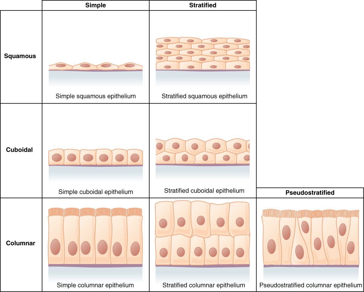
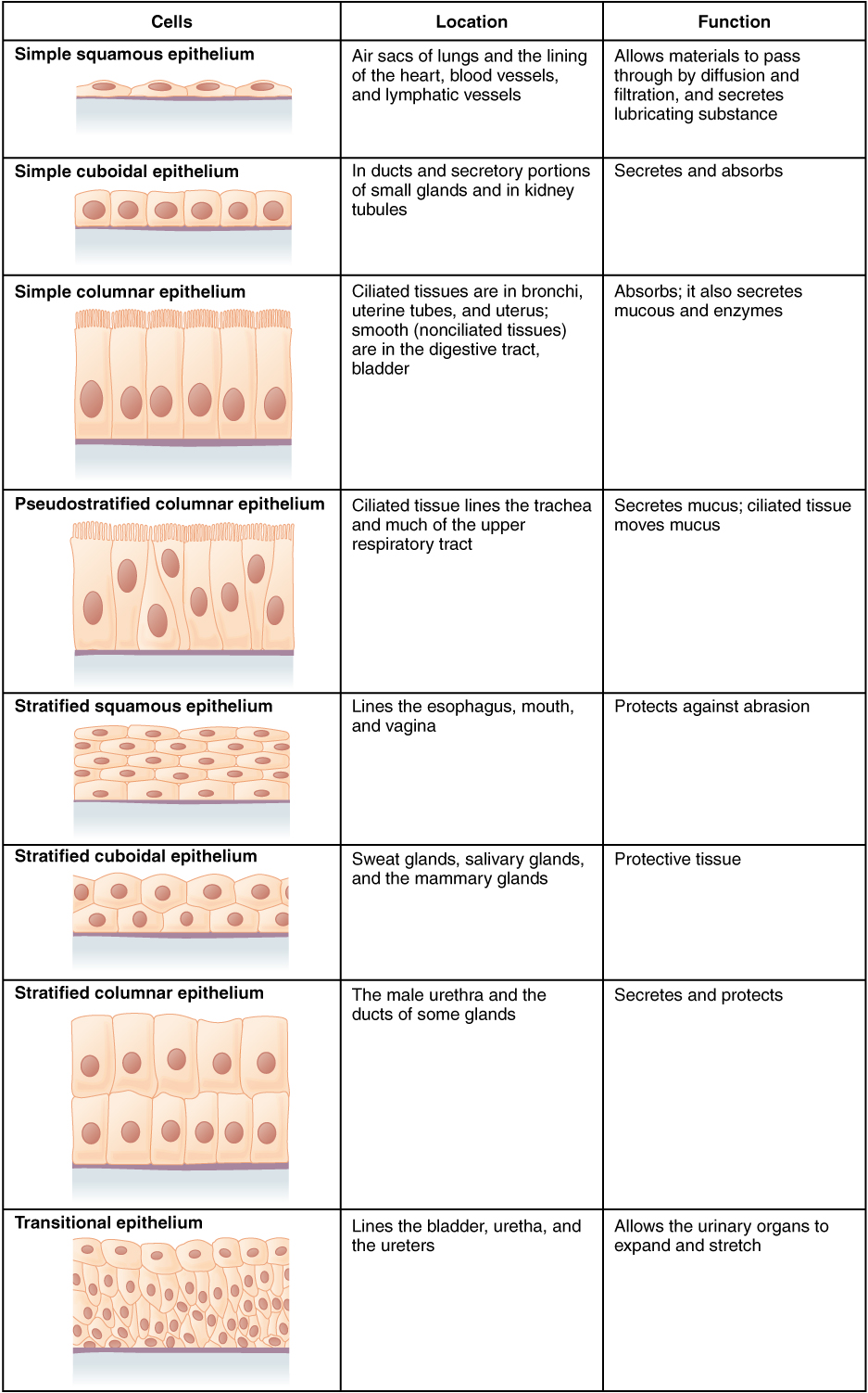
Epithelial tissues are classified co-ordinate to the shape of the cells and number of the prison cell layers formed ((Figure)). Cell shapes tin be squamous (flattened and thin), cuboidal (boxy, as wide every bit information technology is tall), or columnar (rectangular, taller than it is wide). Similarly, the number of cell layers in the tissue tin can exist i—where every cell rests on the basal lamina—which is a simple epithelium, or more than one, which is a stratified epithelium and simply the basal layer of cells rests on the basal lamina. Pseudostratified (pseudo- = "false") describes tissue with a single layer of irregularly shaped cells that requite the appearance of more than ane layer. Transitional describes a grade of specialized stratified epithelium in which the shape of the cells can vary.
Cells of Epithelial Tissue
Simple epithelial tissue is organized every bit a single layer of cells and stratified epithelial tissue is formed by several layers of cells.
Simple Epithelium
The shape of the cells in the single cell layer of elementary epithelium reflects the functioning of those cells. The cells in unproblematic squamous epithelium have the advent of sparse scales. Squamous prison cell nuclei tend to be apartment, horizontal, and elliptical, mirroring the form of the prison cell. The endothelium is the epithelial tissue that lines vessels of the lymphatic and cardiovascular system, and it is made up of a single layer of squamous cells. Simple squamous epithelium, considering of the thinness of the cell, is present where rapid passage of chemical compounds is observed. The alveoli of lungs where gases diffuse, segments of kidney tubules, and the lining of capillaries are likewise made of uncomplicated squamous epithelial tissue. The mesothelium is a simple squamous epithelium that forms the surface layer of the serous membrane that lines torso cavities and internal organs. Its primary function is to provide a smooth and protective surface. Mesothelial cells are squamous epithelial cells that secrete a fluid that lubricates the mesothelium.
In uncomplicated cuboidal epithelium, the nucleus of the box-like cells appears round and is generally located virtually the center of the jail cell. These epithelia are active in the secretion and absorptions of molecules. Unproblematic cuboidal epithelia are observed in the lining of the kidney tubules and in the ducts of glands.
In simple columnar epithelium, the nucleus of the tall column-like cells tends to be elongated and located in the basal stop of the cells. Like the cuboidal epithelia, this epithelium is agile in the absorption and secretion of molecules. Uncomplicated columnar epithelium forms the lining of some sections of the digestive system and parts of the female person reproductive tract. Ciliated columnar epithelium is composed of unproblematic columnar epithelial cells with cilia on their apical surfaces. These epithelial cells are plant in the lining of the fallopian tubes and parts of the respiratory system, where the chirapsia of the cilia helps remove particulate affair.
Pseudostratified columnar epithelium is a type of epithelium that appears to be stratified merely instead consists of a single layer of irregularly shaped and differently sized columnar cells. In pseudostratified epithelium, nuclei of neighboring cells announced at dissimilar levels rather than clustered in the basal end. The arrangement gives the advent of stratification; simply in fact all the cells are in contact with the basal lamina, although some do not attain the apical surface. Pseudostratified columnar epithelium is constitute in the respiratory tract, where some of these cells accept cilia.
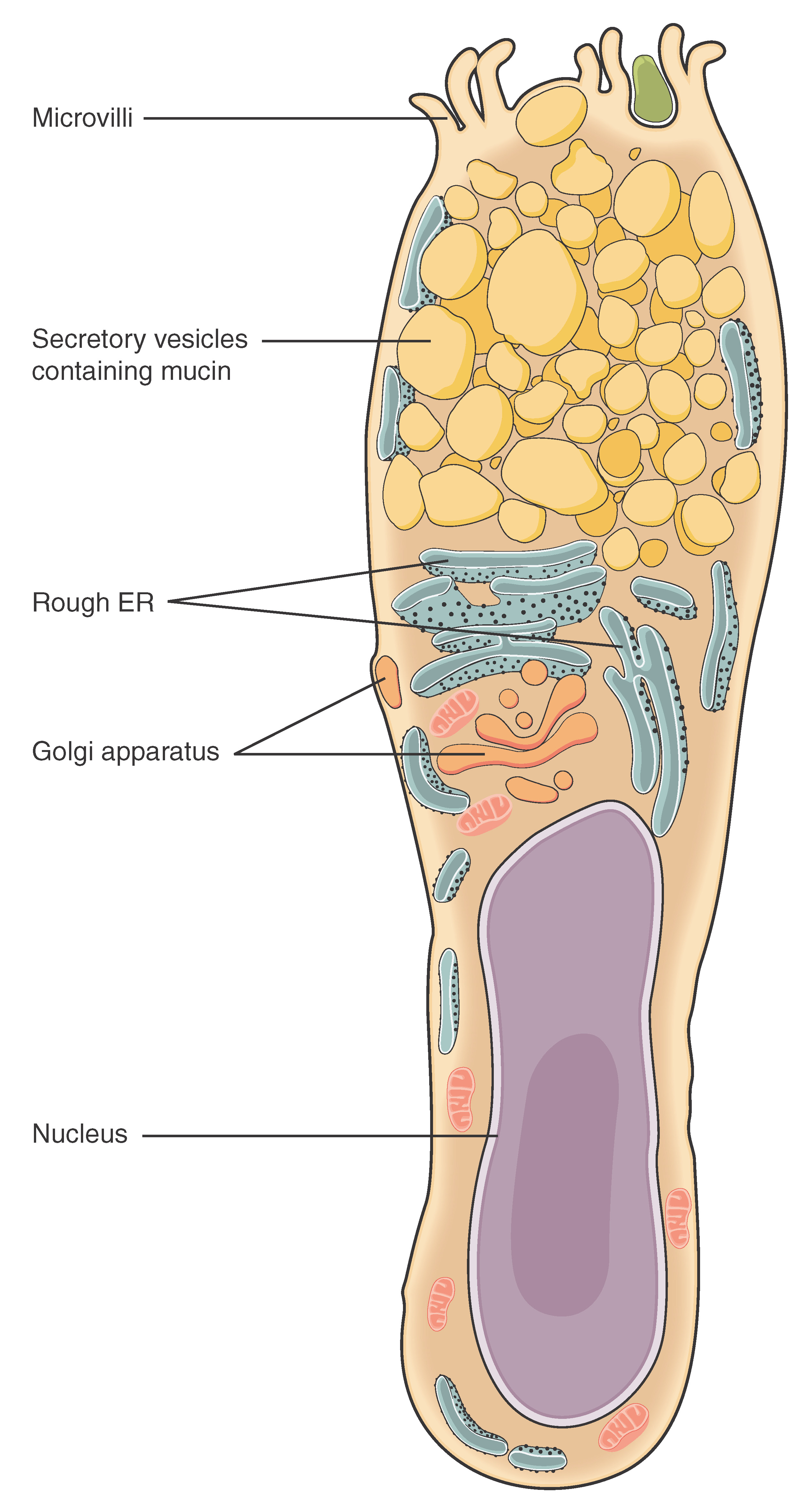
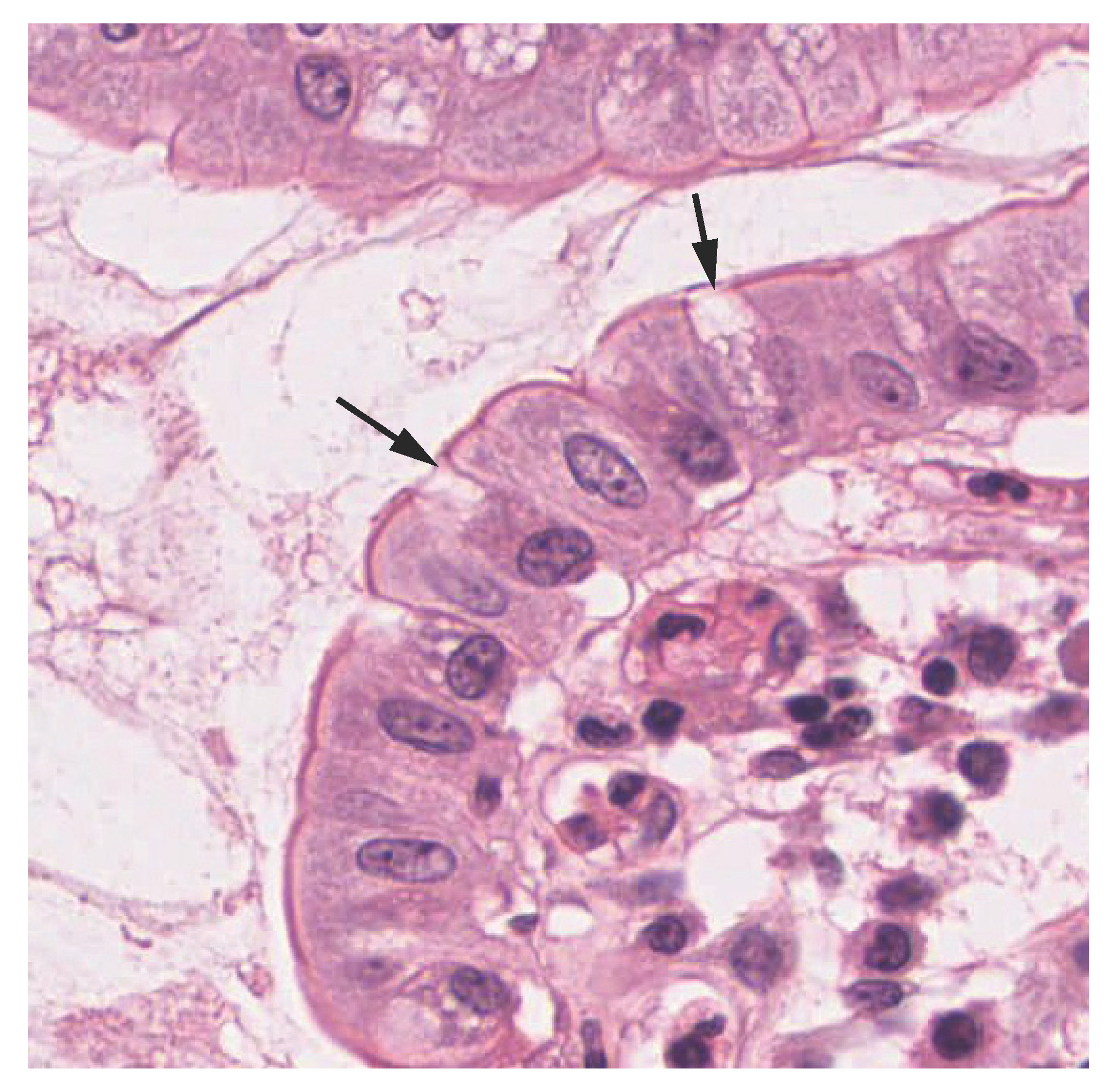
Both simple and pseudostratified columnar epithelia are heterogeneous epithelia considering they include additional types of cells interspersed amidst the epithelial cells. For example, a goblet cell is a mucous-secreting unicellular "gland" interspersed between the columnar epithelial cells of mucous membranes ((Figure)).
Goblet Jail cell
(a) In the lining of the modest intestine, columnar epithelium cells are interspersed with goblet cells. (b) The arrows in this micrograph point to the mucous-secreting goblet cells. LM × 1600. (Micrograph provided by the Regents of University of Michigan Medical School © 2012)
Stratified Epithelium
A stratified epithelium consists of several stacked layers of cells. This epithelium protects against physical and chemical wearable and tear. The stratified epithelium is named by the shape of the most apical layer of cells, closest to the free space. Stratified squamous epithelium is the most common type of stratified epithelium in the human body. The apical cells are squamous, whereas the basal layer contains either columnar or cuboidal cells. The tiptop layer may exist covered with expressionless cells filled with keratin. Mammalian pare is an example of this dry, keratinized, stratified squamous epithelium. The lining of the mouth cavity is an instance of an unkeratinized, stratified squamous epithelium. Stratified cuboidal epithelium and stratified columnar epithelium tin also be establish in certain glands and ducts, just are uncommon in the human trunk.
Another kind of stratified epithelium is transitional epithelium, then-called because of the gradual changes in the shapes of the apical cells as the float fills with urine. Information technology is found just in the urinary system, specifically the ureters and urinary float. When the bladder is empty, this epithelium is convoluted and has cuboidal apical cells with convex, umbrella shaped, apical surfaces. As the bladder fills with urine, this epithelium loses its convolutions and the apical cells transition from cuboidal to squamous. It appears thicker and more multi-layered when the bladder is empty, and more stretched out and less stratified when the float is total and distended. (Figure) summarizes the different categories of epithelial cell tissue cells.
Summary of Epithelial Tissue Cells
Spotter this video to observe out more about the anatomy of epithelial tissues. Where in the torso would one find non-keratinizing stratified squamous epithelium?
Glandular Epithelium
A gland is a structure fabricated upwardly of one or more than cells modified to synthesize and secrete chemical substances. About glands consist of groups of epithelial cells. A gland can be classified equally an endocrine gland, a ductless gland that releases secretions directly into surrounding tissues and fluids (endo- = "inside"), or an exocrine gland whose secretions leave through a duct that opens straight, or indirectly, to the external surroundings (exo- = "exterior").
Endocrine Glands
The secretions of endocrine glands are called hormones. Hormones are released into the interstitial fluid, diffused into the bloodstream, and delivered to targets, in other words, cells that have receptors to bind the hormones. The endocrine organisation is part of a major regulatory arrangement coordinating the regulation and integration of body responses. A few examples of endocrine glands include the inductive pituitary, thymus, adrenal cortex, and gonads.
Exocrine Glands
Exocrine glands release their contents through a duct that leads to the epithelial surface. Mucous, sweat, saliva, and chest milk are all examples of secretions from exocrine glands. They are all discharged through tubular ducts. Secretions into the lumen of the gastrointestinal tract, technically outside of the trunk, are of the exocrine category.
Glandular Structure
Exocrine glands are classified as either unicellular or multicellular. The unicellular glands are scattered single cells, such as goblet cells, found in the mucous membranes of the small and large intestine.
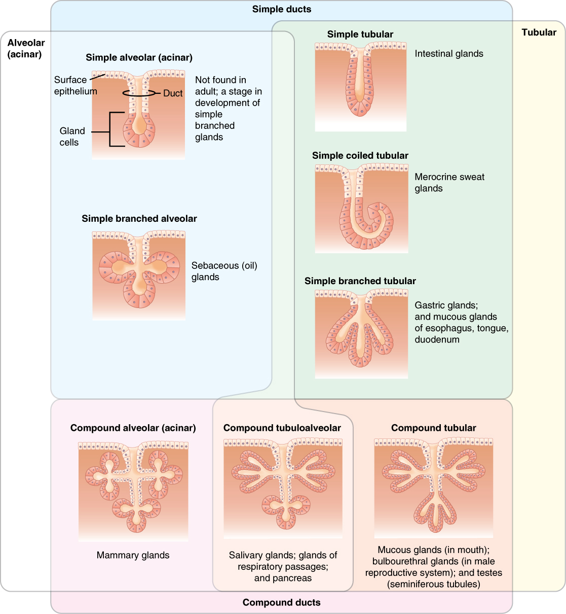
The multicellular exocrine glands known as serous glands develop from uncomplicated epithelium to grade a secretory surface that secretes directly into an inner cavity. These glands line the internal cavities of the abdomen and breast and release their secretions directly into the cavities. Other multicellular exocrine glands release their contents through a tubular duct. The duct is single in a uncomplicated gland but in compound glands is divided into one or more than branches ((Effigy)). In tubular glands, the ducts can be direct or coiled, whereas tubes that form pockets are alveolar (acinar), such as the exocrine portion of the pancreas. Combinations of tubes and pockets are known equally tubuloalveolar (tubuloacinar) compound glands. In a branched gland, a duct is connected to more than than i secretory group of cells.
Types of Exocrine Glands
Exocrine glands are classified by their structure.
Methods and Types of Secretion
Exocrine glands can be classified by their manner of secretion and the nature of the substances released, every bit well every bit past the structure of the glands and shape of ducts ((Effigy)). Merocrine secretion is the nearly common type of exocrine secretion. The secretions are enclosed in vesicles that move to the apical surface of the jail cell where the contents are released past exocytosis. For example, watery mucous containing the glycoprotein mucin, a lubricant that offers some pathogen protection is a merocrine secretion. The eccrine glands that produce and secrete sweat are another case.
Modes of Glandular Secretion
(a) In merocrine secretion, the jail cell remains intact. (b) In apocrine secretion, the apical portion of the cell is released, as well. (c) In holocrine secretion, the jail cell is destroyed as it releases its product and the cell itself becomes function of the secretion.
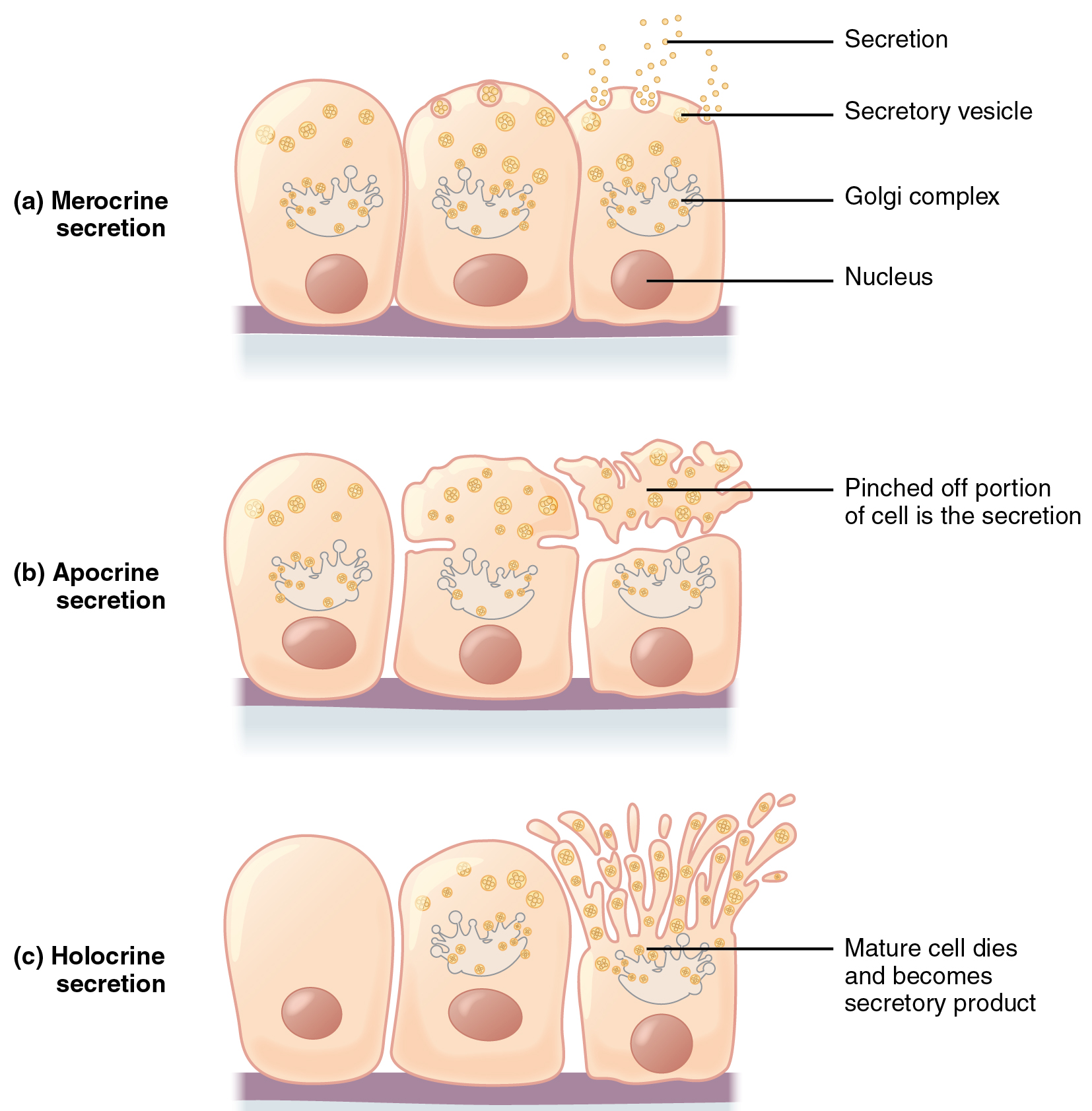
Apocrine secretion accumulates near the upmost portion of the cell. That portion of the prison cell and its secretory contents compression off from the cell and are released. Apocrine sweat glands in the axillary and genital areas release fatty secretions that local bacteria intermission downwardly; this causes body odor. Both merocrine and apocrine glands go along to produce and secrete their contents with little damage caused to the jail cell considering the nucleus and golgi regions remain intact after secretion.
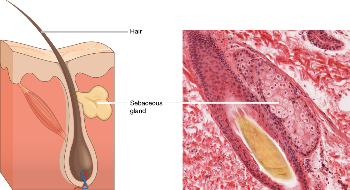
In contrast, the process of holocrine secretion involves the rupture and destruction of the entire gland cell. The jail cell accumulates its secretory products and releases them merely when information technology bursts. New gland cells differentiate from cells in the surrounding tissue to supersede those lost past secretion. The sebaceous glands that produce the oils on the peel and hair are holocrine glands/cells ((Figure)).
Sebaceous Glands
These glands secrete oils that lubricate and protect the skin. They are holocrine glands and they are destroyed after releasing their contents. New glandular cells form to replace the cells that are lost. LM × 400. (Micrograph provided by the Regents of University of Michigan Medical School © 2012)
Glands are also named after the products they produce. The serous gland produces watery, blood-plasma-like secretions rich in enzymes such every bit alpha amylase, whereas the mucous gland releases watery to viscous products rich in the glycoprotein mucin. Both serous and mucous glands are common in the salivary glands of the oral cavity. Mixed exocrine glands contain both serous and mucous glands and release both types of secretions.
Chapter Review
In epithelial tissue, cells are closely packed with piddling or no extracellular matrix except for the basal lamina that separates the epithelium from underlying tissue. The main functions of epithelia are protection from the surroundings, coverage, secretion and excretion, absorption, and filtration. Cells are spring together by tight junctions that form an impermeable barrier. They can also be connected past gap junctions, which allow gratis exchange of soluble molecules between cells, and anchoring junctions, which adhere cell to cell or cell to matrix. The different types of epithelial tissues are characterized by their cellular shapes and arrangements: squamous, cuboidal, or columnar epithelia. Single cell layers form simple epithelia, whereas stacked cells form stratified epithelia. Very few capillaries penetrate these tissues.
Glands are secretory tissues and organs that are derived from epithelial tissues. Exocrine glands release their products through ducts. Endocrine glands secrete hormones directly into the interstitial fluid and blood stream. Glands are classified both according to the blazon of secretion and by their construction. Merocrine glands secrete products as they are synthesized. Apocrine glands release secretions past pinching off the apical portion of the cell, whereas holocrine gland cells store their secretions until they rupture and release their contents. In this case, the cell becomes role of the secretion.
Interactive Link Questions
Sentinel this video to find out more about the anatomy of epithelial tissues. Where in the torso would ane find non-keratinizing stratified squamous epithelium?
The inside of the mouth, esophagus, vaginal canal, and anus.
Review Questions
In observing epithelial cells under a microscope, the cells are bundled in a single layer and wait alpine and narrow, and the nucleus is located close to the basal side of the prison cell. The specimen is what blazon of epithelial tissue?
- columnar
- stratified
- squamous
- transitional
Which of the post-obit is the epithelial tissue that lines the interior of blood vessels?
- columnar
- pseudostratified
- simple squamous
- transitional
Which type of epithelial tissue specializes in moving particles beyond its surface and is found in airways and lining of the oviduct?
- transitional
- stratified columnar
- pseudostratified ciliated columnar
- stratified squamous
The ________ exocrine gland stores its secretion until the glandular prison cell ruptures, whereas the ________ gland releases its apical region and reforms.
- holocrine; apocrine
- eccrine; endocrine
- apocrine; holocrine
- eccrine; apocrine
Critical Thinking Questions
The construction of a tissue ordinarily is optimized for its function. Describe how the structure of the mucosa and its cells friction match its role of nutrient assimilation.
The mucosa of the intestine is highly folded, increasing the surface area for nutrient assimilation. A greater surface surface area for assimilation allows more nutrients to be absorbed per unit time. In add-on, the food-absorbing cells of the mucosa have finger-like projections called microvilli that further increase the expanse for nutrient assimilation.
Glossary
- anchoring junction
- mechanically attaches adjacent cells to each other or to the basement membrane
- apical
- that part of a cell or tissue which, in general, faces an open space
- apocrine secretion
- release of a substance along with the apical portion of the cell
- basal lamina
- thin extracellular layer that lies underneath epithelial cells and separates them from other tissues
- basement membrane
- in epithelial tissue, a thin layer of gristly textile that anchors the epithelial tissue to the underlying connective tissue; made upward of the basal lamina and reticular lamina
- jail cell junction
- point of cell-to-cell contact that connects one cell to another in a tissue
- endocrine gland
- groups of cells that release chemical signals into the intercellular fluid to exist picked up and transported to their target organs by blood
- endothelium
- tissue that lines vessels of the lymphatic and cardiovascular system, made up of a simple squamous epithelium
- exocrine gland
- group of epithelial cells that secrete substances through ducts that open up to the pare or to internal torso surfaces that pb to the exterior of the trunk
- gap junction
- allows cytoplasmic communications to occur between cells
- goblet cell
- unicellular gland found in columnar epithelium that secretes mucous
- holocrine secretion
- release of a substance acquired past the rupture of a gland cell, which becomes part of the secretion
- merocrine secretion
- release of a substance from a gland via exocytosis
- mesothelium
- simple squamous epithelial tissue which covers the major body cavities and is the epithelial portion of serous membranes
- mucous gland
- group of cells that secrete mucous, a thick, slippery substance that keeps tissues moist and acts as a lubricant
- pseudostratified columnar epithelium
- tissue that consists of a unmarried layer of irregularly shaped and sized cells that give the appearance of multiple layers; found in ducts of certain glands and the upper respiratory tract
- reticular lamina
- matrix containing collagen and elastin secreted by connective tissue; a component of the basement membrane
- serous gland
- group of cells within the serous membrane that secrete a lubricating substance onto the surface
- uncomplicated columnar epithelium
- tissue that consists of a single layer of column-like cells; promotes secretion and assimilation in tissues and organs
- simple cuboidal epithelium
- tissue that consists of a single layer of cube-shaped cells; promotes secretion and absorption in ducts and tubules
- uncomplicated squamous epithelium
- tissue that consists of a single layer of flat scale-like cells; promotes diffusion and filtration beyond surface
- stratified columnar epithelium
- tissue that consists of ii or more layers of column-similar cells, contains glands and is found in some ducts
- stratified cuboidal epithelium
- tissue that consists of two or more layers of cube-shaped cells, plant in some ducts
- stratified squamous epithelium
- tissue that consists of multiple layers of cells with the well-nigh apical existence flat scale-like cells; protects surfaces from abrasion
- tight junction
- forms an impermeable barrier between cells
- transitional epithelium
- class of stratified epithelium found in the urinary tract, characterized by an apical layer of cells that modify shape in response to the presence of urine
Source: https://opentextbc.ca/anatomyandphysiologyopenstax/chapter/epithelial-tissue/
Posted by: torrezandessaint.blogspot.com
0 Response to "Where In The Body Would You Be Most Likely To Find Secretory Cells Shaped Like Dice?"
Post a Comment